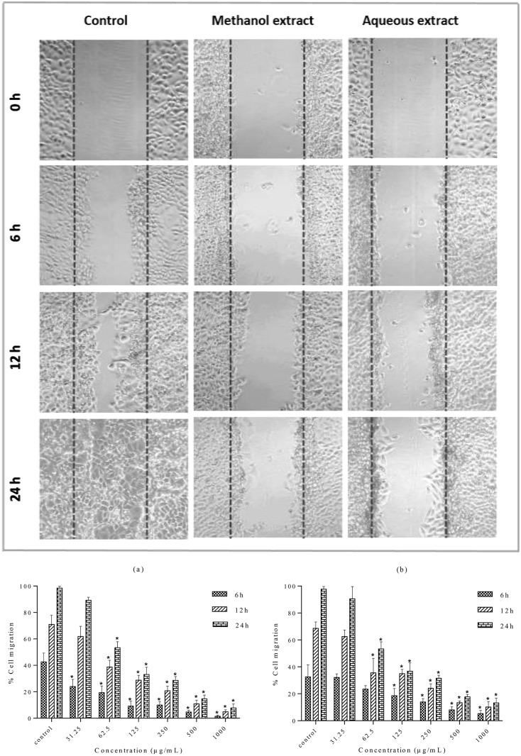
Fig 9. Effect of S. ferruginea methanolic and aqueous extracts on the cell migration of MDA-MB-231 cells.
(A) Wound closure ability of treated MDA-MB-231 cells after creation of scratch wound in control and treated well. The images of wounded extract-treated MDA-MB-231 cell monolayers captured using an inverted phase-contrast microscope at different time intervals (0, 6, 12, and 24 h) are shown. (B) Quantitative measurement of MDA-MB-231 cell migration after treatment with methanolic (a) and aqueous (b) extracts at different concentrations (31.25–1000 μg/mL). Wound closure rates were quantitatively analyzed by calculating the difference between wound width at 0, 6, and 12 or 24 h of extract-treated and control cells. Results are expressed as percentage of cell migration. Data are means ± SD of triplicate in three independent experiments. * indicates statistically significant difference from their respective untreated control (one-way ANOVA, P < 0.05).

