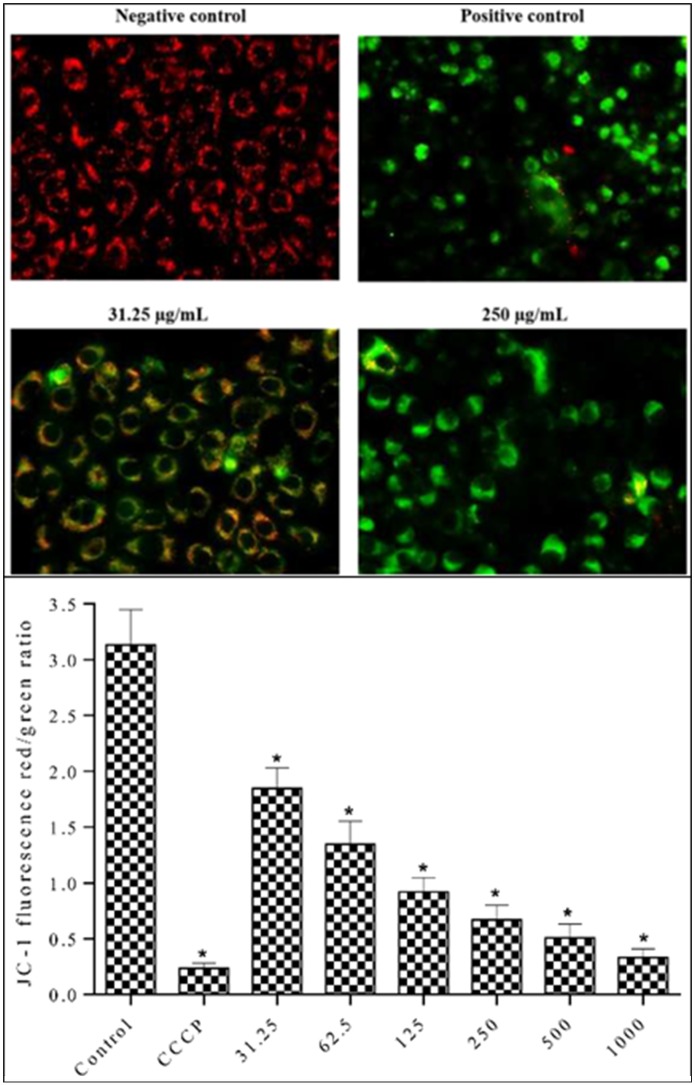
Fig 11. Effect of methanolic extract on MMP in MDA-MB-231 cells using JC-1 fluorescence dye.
A. Methanolic extract induced MMP depolarization in MDA-MB-231 cells. The cells were treated with extracts at 31.25 μg/mL and 250 μg/mL and with positive control (50 μM CCCP) for 12 h. Cells were imaged with an inverted fluorescence microscope (Zeiss Axiovert A) at 40x magnification. The emitted green fluorescence indicates MMP depolarization, which is an early event in apoptosis. B. Relative quantification of MMP (ΔΨm) in MDA-MB-231 cells. Cells were treated with different concentrations of methanolic extract and positive control (50 μM CCCP) for 12 h. Methanolic extract disrupts MMP (ΔΨm). Data are means ± SD of triplicate in three independent experiments. * indicates statistically significant difference from corresponding controls (one-way ANOVA, P < 0.05).

