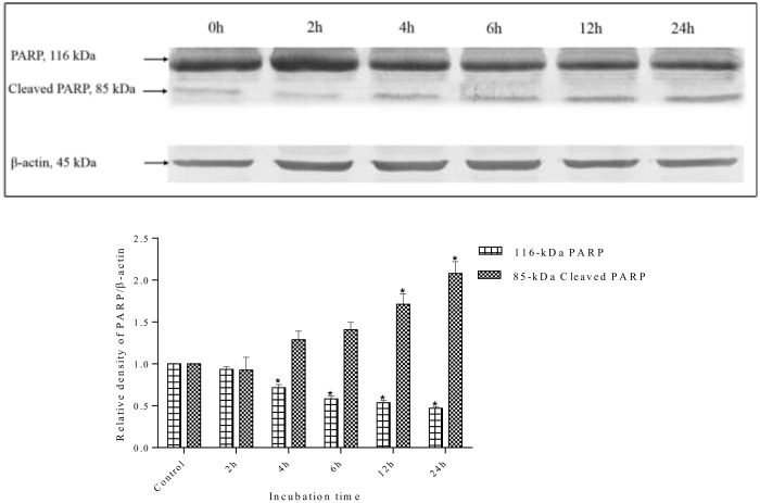
Fig 16. Western blot analysis of PARP protein in MDA-MB-231 cells.
MDA-MB-231 cells were treated with S. ferruginea methanolic extract at IC50 for indicated times. Control cells were treated with 0.1% DMSO. β-Actin was used as loading control. The PARP protein (116-kDa) was cleaved into its signature 85-kDa fragment, a marker of apoptosis, after treatment with the methanolic extract. The densitometric-intensity data are presented as means ± SEM of triplicate in three independent experiments. * indicates statistically significant difference from control (one-way ANOVA, P < 0.05).

