
Fig. 2.
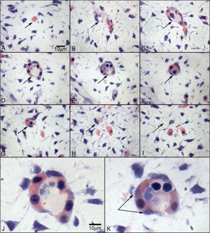
Wright’s Giemsa staining of 2.5-μm plastic sections showing an island of cells in the choroid at 6 WG. Serial sections (A–I) confirm that the structure is isolated and separated from any blood vessels at 6 WG. The arrow shows the same position in each panel. J,K: High-magnification photos of D and E, respectively. Wright’s Giemsa staining shows acidophilic cytoplasm indicative of hemoglobin in pink and basophilic nuclei in blue. One cell outside of structure (double arrow in K) shows the changing of cytoplasmic staining pattern from acidophilic into basophilic, which suggests that the cell has both the characteristics of hematopoietic cells and endothelial cells. In J, the erythroblasts appear to form the lumen.