
Fig. 4.
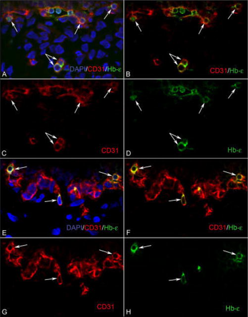
The developing choroidal vasculature contains CD31/Hb-∈ double positive cells. A–D: At 6 WG, clusters of CD31/Hb-∈ (red/green) positive cells (arrows) are visible in the choriocapillaris layer, whereas in the choroidal stroma, there are isolated cells (double arrow) that are also double labeled. E–H: At 7 WG, there are isolated cells (arrows) that are CD31/Hb-∈ positive, which appear attached to the choriocapillaris. These CD31/Hb-∈-positive cells have characteristics of both hematopoietic and endothelial cells. B and F are merged images of the single color images C, D, and G, H, respectively. A and E are images showing nuclear counter staining with DAPI (blue) merged with B and F, respectively.