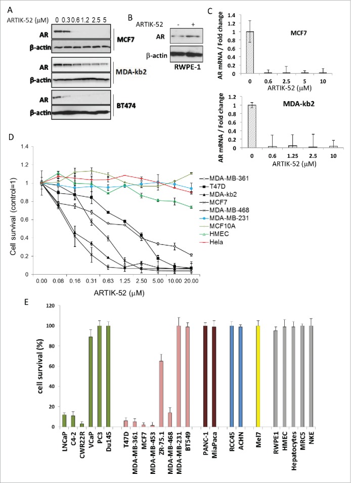
Figure 2.

ARTIK-52 treatment causes reduction of AR levels and toxicity in AR expressing tumor cells. A. AR protein detected by western blotting in BC cells treated with indicated concentrations of ARTIK-52 for 24 h. B. Western blotting of extracts of normal prostate cells, RWPE-1, treated with 1 µM of ARTIK-52 for 24 h, stained with the indicated antibodies. C. AR mRNA levels detected using qPCR in BC cells treated with indicated concentrations of ARTIK-52 for 24 h. Error bars represent standard deviation between 3 replicates within one experiment. D. Survival of cells maintained with different concentrations of ARTIK-52 for 72 hours. Black lines correspond to AR positive tumor cell lines, colored lines – to AR negative tumor cell lines and non-tumor cells. Error bars represent standard deviation between 3 replicates in at least 2 different experiments E. Survival of cells treated with 5 µM of ARTIK-52 for 72 h. Untreated cells were taken as 100%. Green bars correspond to prostate cancer cells, pink – breast cancer cells, brown – pancreatic cancer, blue – renal cancer, yellow – melanoma, gray – non-tumor cells: RPWE-1 – prostate epithelial cell line, HMEC – primary mammary epithelia, MRC5 – diploid fibroblasts, NKE – kidney epithelial cell line. Error bars represent standard deviation between 3 replicates in at least 2 different experiments.
