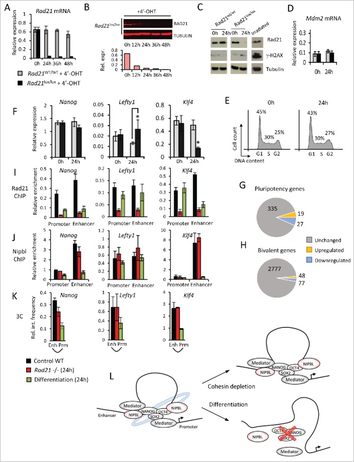
Figure 2.

Acute cohesin depletion is compatible with pluripotency gene expression and enhancer-promoter interactions. (A-E) Time course of Rad21 mRNA (A) and RAD21 protein depletion (B) induced by 4′-OHT-mediated activation of ERt2Cre in ERT2Cre-Rad21lox/lox ES cells (100nM 4′-OHT). Acute cohesin depletion did not result in significant DNA damage as indicated by phosphorylation of H2AX (γ-H2AX), irradiated ES cells were used as positive control (C); upregulation of the p53 target gene Mdm2 (D) or cell cycle arrest (E). (F) Quantitative RT-PCR analysis of selected pluripotency genes in ES cells before (0h) and after acute cohesin depletion (24h). (G, H) Genome-wide expression analysis of pluripotency genes (G) and bivalent genes (H) in acutely cohesin-depleted ES cells at 24 hours. (I,J) ChIP for RAD21 (I) and NIPBL (J) at the promoters and enhancers of Nanog, Lefty1 and Klf4 in control ES cells (black), 24h Rad21-deleted ES cells (red) and differentiating ES cells (green). (K) Chromosome conformation capture (3C) assays for promoter-enhancer interactions at Nanog, Lefty1 and Klf4 in control ES cells (black), 24h Rad21-deleted ES cells (red) and differentiating ES cells (green) (L) Enhancer-promoter interactions at pluripotency loci in ES cells (left) are maintained after acute cohesin depletion (right, top) but lost during ES cell differentiation (right, bottom).
