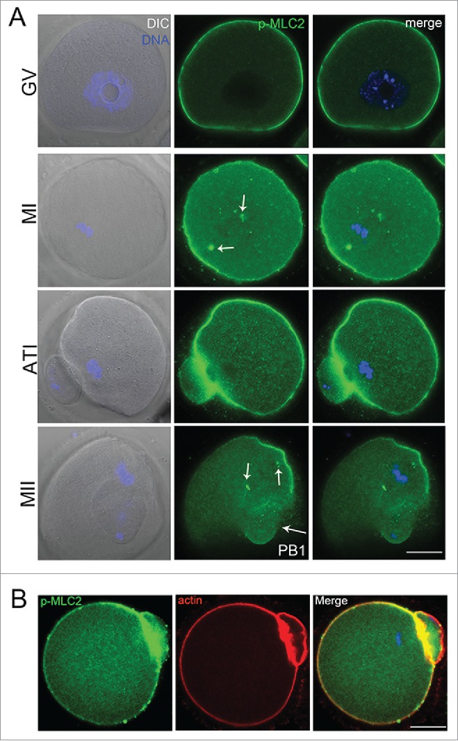
Figure 1.

(A) Confocal imaging analysis of the p-MLC2 localization during mouse oocyte meiotic maturation. Oocyte at GV, metaphase I (MI), telophase (ATI) and metaphase II stages were immunolabeled with p-MLC2 antibody. (B) p-MLC2 was co-localized with actin in oocyte membrane. Arrows showed that the localization of p-MLC2 at the spindle poles. Blue: chromatin; Green: p-MLC2; Red: actin. Bar = 20 μm.
