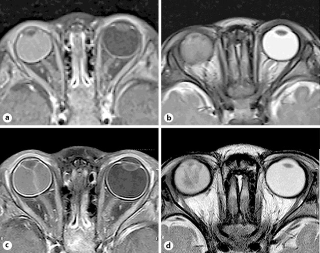
Fig. 3.

MRIs taken at two different periods. T1 (a) and T2 (b) images taken 3 months before the first visit show a hyperintense lesion and a slightly hypointense lesion in the subretinal space, respectively. Both T1 (c) and T2 (d) images taken at the first visit show hyperintense lesions in the subretinal space. Tumor structure is not observed in the eye.
