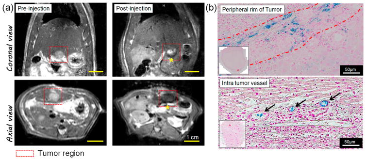
Figure 5.
MRI-monitored hepatic IA transcatheter infusion of pH-DENs in orthotopic HCC rat model. (a) In vivo T2-weighted MR images acquired before and after transcatheter infusion of pH-DENs. Intrahepatic deposition of pH-DENs is depicted as regions of signal loss within the T2-weighted images postinfusion (yellow asterisk). (b) Prussian blue staining of treated tumor-bearing liver tissues 24 h post-IA injection. (Top) Peripheral rim and (bottom) intratumor vessels at edge of tumor.

