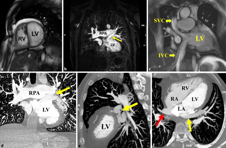
Fig. 1.
a CMR short-axis steady-state free precession (SSFP) coronal image showing hypertrabeculated systemic RV with dilated LV. b CMR angiography revealing the left lower PV draining superiorly to a larger common left-side PV which in turn connects to the superior aspect of the left atrium (arrow). Right pulmonary artery (RPA) is dilated. c CMR SSFP coronal image revealing patent superior (SVC) and inferior vena cava (IVC) limbs of the Senning baffle. d CCT maximal intensity projection coronal image showing a tortuous varix of the left lower PV draining superiorly to a larger common left-side PV which in turn connects to the superior aspect of the left atrium. RPA is dilated. e CCT sagittal view revealing the tortuous varix (arrow). f CCT axial view showing the site of anastomosis between the left atrium and anomalous PV connection (yellow arrow). Patent pulmonary venous baffle was noted (red arrow)

