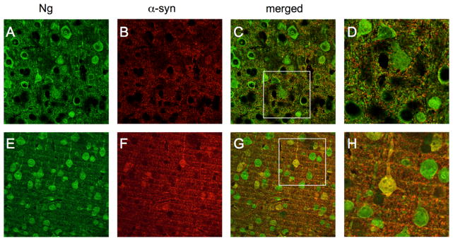
Fig. 2.
Neurogranin interaction with α-synuclein in Tg mouse brain. Immunohistochemical neurogranin expression in nonTg mouse brains (green, (A)) when compared with α-syn (red, (B)) revealed no colocolization among cell bodies ((C) and (D)) but possible colocalization in the synapse. In Tg mouse brains, neurogranin (E) and α-syn (F) showed obvious yellow colocalization in cell bodies and synapes ((G) and (H)). (For interpretation of the references to color in this figure legend, the reader is referred to the web version of this article.)

