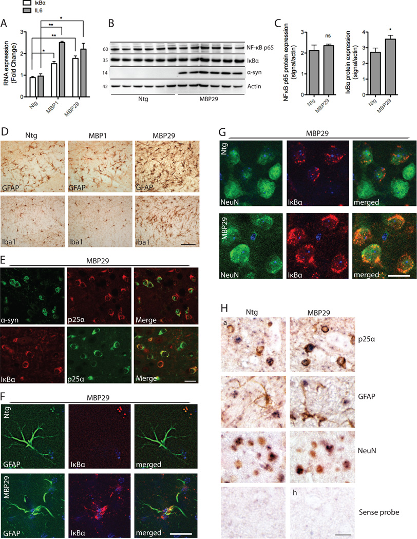
Figure 4. IκBα expression in MBP-hα-syn transgenic mice.
A, IκBα and IL6 mRNA from total brain extracts of MBP-hα-syn tg (line 1 and 29) and ntg mice (n=6; age=3 months) were analyzed by real-time qPCR. Fold changes were determined from triplicate measurements and normalized to mouse GAPDH. Bars represent mean ± S.D. from three independent experiments. IκBα and IL6 mRNA are significantly increased in both line 1 and 29 as compared to ntg mice (*p < 0.05, **p < 0.01). B, IκBα and NF-κB p65 protein levels in line 29 mouse brain homogenates were analyzed by western blot analysis. β-actin is included as a loading control. Representative blots from n=6 of each group are shown. C, Quantitative analysis of the levels of IκBα and NF-κB p65 demonstrates a significant increase in IκBα expression in tg mice compared to ntg controls (*p < 0.05) whereas there was no difference in NF-κB p65 levels between groups. D, Immunohistochemical analysis of corpus callosum with antibodies against GFAP and Iba1 shows that MBP-hα-syn line 29 (3 months old) displays severe astrogliosis and microgliosis in corpus callosum as compared to a ntg control (a,d). There was no difference in the expression of these proteins between the intermediate-expresser line 1 and the ntg control. Scale bar, 50 µm. E, Double labeling immunofluorescence microscopy of corpus callosum reveals colocalization of α-syn (green) and p25α (red) and of IκBα (red) and p25α (green) in corpus callosum of 3 months old line 29 mice. Scale bar, 30 µm. F–G, Double labeling studies for IκBα in neocortex of line 29 mice. Sections were double labeled with antibodies against the astroglial marker GFAP (F) and the neuronal marker NeuN (G) and IκBα. Scale bar, 10 µm. F, There was IκBα immunostaining of astrocytes in line 29 mice compared to ntg controls. G, In ntg mice occasional IκBα immunoreactivity was observed. In the MBP-α-syn tg mice there was an increase in IκBα immunolabeling. H, To validate the cell type-specific IκBα mRNA expression, in situ hybridization with a probe against IκBα (blue) and co-immunolabeling with markers for oligodendrocytes (p25α) and astroglial (GFAP) in corpus callosum, and neuronal (NeuN) antibodies (brown) in neocortex was conducted in 3 months old ntg and line 29 mice. In situ hybridization with sense probe showed no signal. Scale bar, 30 µm.

