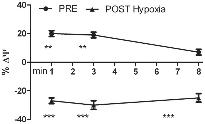
Figure 5.

Kinetics of the percentage variation of transmembrane mitochondrial potential (ΔΨ) of PRE- and POST-Hypoxia differentiated cells after stimulation with 100 nM H2O2. The fluorescence of control cells for both PRE- and POST-Hypoxia conditions, were fixed as 0% and not showed in the graph. The PRE-Hypoxia cells after H2O2 stimulation showed a significant hyperpolarization of the transmembrane mitochondrial potentials while the POST-Hypoxia cells a significant depolarization. **p ≤ 0.01, ***p ≤ 0.0001 vs. no H2O2-treated cells.
