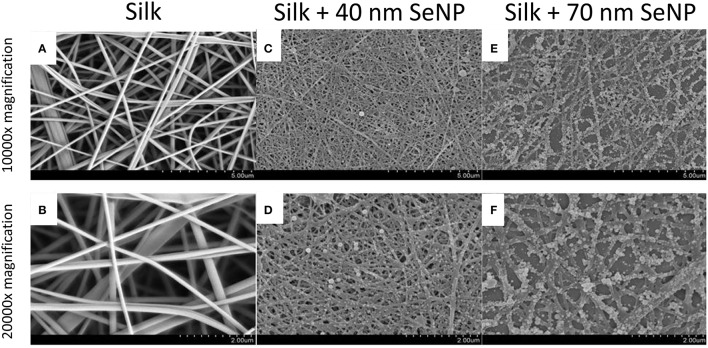
Figure 1.
Scanning electron microscopy (SEM) images of the electrospun silk scaffolds at 10,000x (A,C,E) and 20,000x (B,D,F) with 5 and 2 μm scale bars respectively. The silk scaffolds without selenium nanoparticles are shown in panels (A,B); with 40 nm selenium nanoparticle in panels (C,D); and with 70 nm selenium nanoparticles in panels (E,F).

