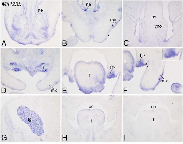
Figure 3.
Expression of Mir23b in mouse facial structures at E12.5. ISH analysis in frozen frontal sections through the head of E12.5 mouse embryos. (A–C) Mir23b is moderately expressed in the nasal epithelium epithelium (ne), mystacial vibrissae (mv), nasal septum (ns), and vomeronasal organ (vo). (D–F) Mir23b is also expressed in the upper incisors (i), tongue (t), maxillary epithelium (te), and palatal shelves (ps). The arrow in (F) denotes the epithelium where Mir23b expression begins. (G–I) More posteriorly, expression is observed in the trigeminal ganglion (tg; G), posterior tongue (t; H,I). i, incisor; mx, maxilla; oc, oral cavity.

