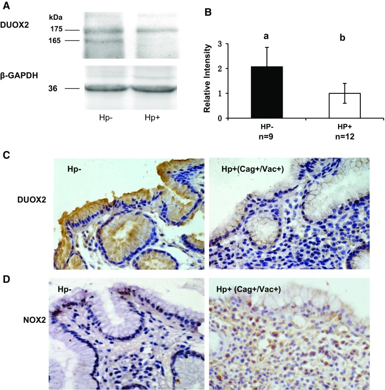
Fig. 3.
Comparison of DUOX protein expression in gastric mucosa of Hp− and Hp+ patients. a Western blot of DUOX and GAPDH from a representative Hp− and Hp+ patients. The expected m.w. of DUOX is 175 kDa, and the Hp− has an additional 165-kDa band recognized by anti-DUOX2 Ab (Abcam, which cannot distinguish DUOX2 from DUOX1). b Bar graph of DUOX protein quantified from the Western blots analyzed from 9 Hp− and 12 Hp+ patients. The error bar is SEM; the different letters above the columns indicate that the means are different, where a > b (P < 0.05). c IHC stained gastric mucosa from Hp− and Hp+ (CagA+/VacA) patients. Hp+ gastric mucosa has more infiltrating cells and weaker DUOX staining (brown color; the original magnification is ×400). d That there is higher NOX2 staining in inflammatory cells in the Hp+ (CagA+/VacA) gastric mucosa than that in Hp− (brown color; the original magnification is ×400)

