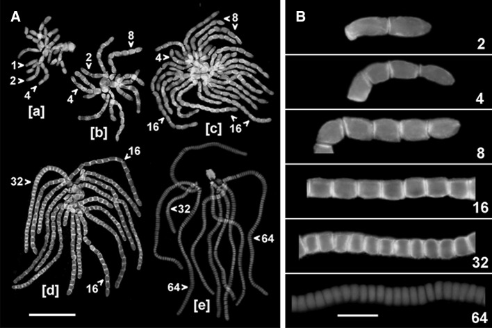
Fig. 2.
Immunfluorescence of PIN2-LPs during development of antheridial filaments in C. vulgaris. Selected filaments are indicated (arrowheads) by the number of component cells (spermatids). a Rosettes of filaments at early [a], middle [b, c], and late [d] stages of the proliferative period and at the onset of differentiation into mature sperm cells [e]. Nuclear DNA stained blue with DAPI. Scale bar 75 μm. b Antheridial filaments at 2- to 64-cell stages (numbers) showing differences in the intensity of PIN2-LPs immunofluorescence localized to the transverse cell wall regions between adjacent spermatids. Scale bar 20 μm

