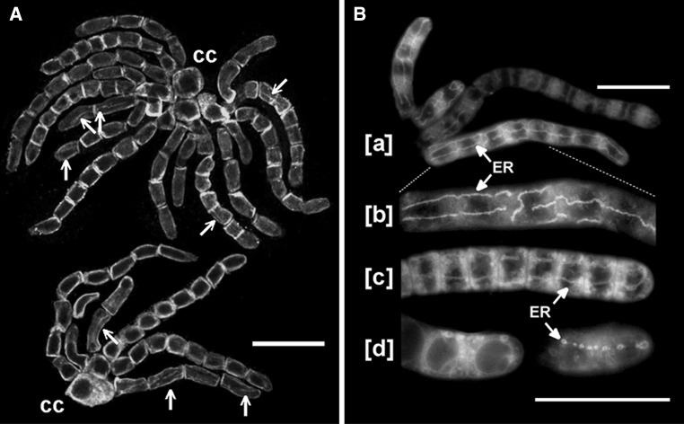
Fig. 6.
Immunofluorescence labeling of PIN2-LPs reveals tubular structures which may correspond to DiOC6(3)-stained cisterns of endoplasmic reticulum. a Confocal images of PIN2-LPs in two rosettes of two-, four-, and eight-celled antheridial filaments attached to a complex of capitular cells (cc); spermatids displaying tubular structures are indicated (arrows). Scale bar 20 μm. b Live antheridial filaments stained fluorescently with DiOC6(3) under the conventional fluorescence microscope. [a] Portion of a rosette of filaments with cisterns of endoplasmic reticulum (ER). Scale bar 25 μm. [b] Enlarged fragment of the eight-celled antheridial filament shown in [a]. [c] Portion of antheridial filament at late stage of the proliferative period showing DiOC6(3)-stained ER cisterns. [d] Apical cells of an antheridial filament at different focal planes displaying nuclear envelope membranes (left) and a row of vesicular ER compartments (right). Scale bar for [b–d] 25 μm

