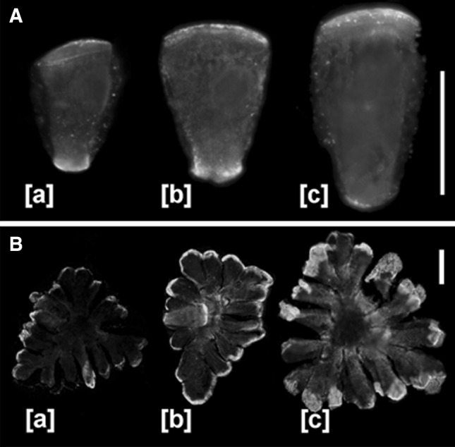
Fig. 7.

Immunofluorescence of PIN2-LPs in manubria and shield cells. a Localization of PIN2-LPs in manubria depends on the developmental period of an antheridium. During the early stages of the proliferative period [a], PIN2-LPs are confined mostly to the shorter cell wall connected with the central complex of capitular cells, while at later stages [b, c] the dominant polarization of PIN2-LPs changes towards the longer transverse cell wall adjoined to the shield cell. Scale bar 40 μm. b The quantity of PIN2-LPs localized to the multi-lobed outer cell walls of the shield cell gradually increases during successive phases of antheridial development [a, b], reaching maximum at late stages of spermatogenesis [c]. In [b], manubrium attached to the central part of the shield cell is visible. Scale bar 40 μm
