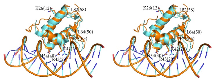
Figure 4.
Stereo superposition between the wHTH domains of GabR from Bacillus subtilis and FadR from E. coli. Stereo superposition between the wHTH domains of GabR from Bacillus subtilis (orange cartoon, PDB code 4TV7) and of FadR from E. coli (cyan, PDB code 1H9T). GabR and FadR residues discussed in the text are displayed as sticks. Labels denote GabR residues with amino acid single letter code. Numbering refers to GabR structure and to Figure 3 (in parentheses). Table 2 reports the correspondence between the residues and their numbering. In particular, Arg29 corresponds to GabR Arg43 and FadR Arg35. DNA phosphate backbone is displayed as orange wire while bases are depicted with cyan and blue sticks.

