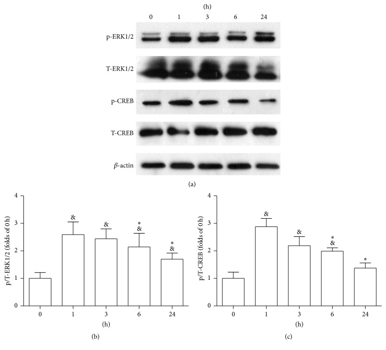
Figure 6.
(a) Western blotting was performed to evaluate the expression levels of p-ERK1/2 and p-CREB. Brain tissues were extracted at 0 h, 1 h, 3 h, 6 h, and 24 h after MCAO. The phosphorylation of ERK1/2 (b) and CREB (c) peaked at 1 h. Data are expressed as mean ± SD; n = 3 rats/group (& P < 0.05 versus 0 h; ∗ P < 0.05 versus 1 h).

