Figure 1.
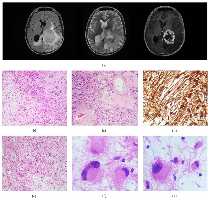
Magnetic resonance imaging scans/histopathological findings. (a) Postcontrast T1 (right) and T2 weighted (center) sequences demonstrate a cystic, avidly enhancing mass, while the fluid-attenuated inversion recovery (FLAIR) sequence (left) shows enlargement of the cornu occipitale sinister with surrounding white matter oedema, consistent with ventricular entrapment; this might as well explain the blurred vision due to affection of the geniculocalcarine tract. (b) Transition zone, in a spindle cell neoplastic population, from an Antony A pattern (left) to an Antony B area (right). H&E. (c) High magnification photomicrograph showing intratumoural hyalinised vessels. H&E. (d) Reticular fibers stain. A pericellular reticulin frame, trademark of neurilemmal phenotype, is noticeable. (e) Antony B field with a small population of neoplastic, plump, gemistocyte-like cells. H&E. ((f), (g)) High magnification photomicrographs of the cells shown in (e): voluminous cells with eccentric nuclei and senescent atypia are evident.

