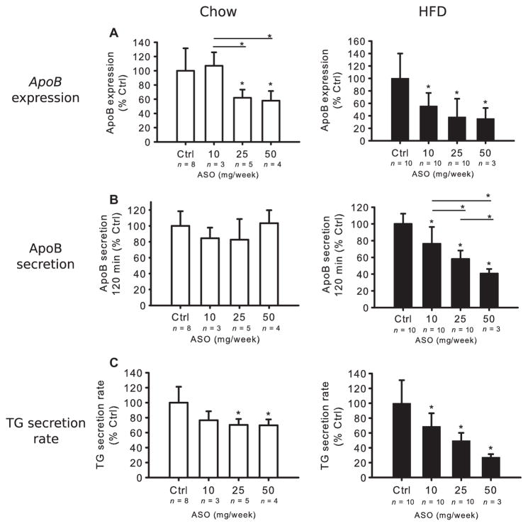
Fig. 3. Effects of ApoB ASO treatment on apoB and TG secretion in Apobec-1 knockout mice.
Chow-and HFD-fed mice were treated for 3 weeks with ApoB ASO. (A) ApoB mRNA expression was determined using qPCR of livers from both chow and HFD mice and normalized to liver β-actin mRNA. In each diet group, irrelevant, control (Ctrl) ASO-treated mRNA levels were set as 100%. (B) Mice were fasted 4 hours and injected with Tyloxapol and [35S]methionine intravenously. Plasma 35S-labeled apoB levels in CPM were determined in samples obtained at 120 min after the start of the study. ApoB radioactivity in mice receiving the irrelevant control ASO was set as 100%. (C) Blood samples were collected at 0, 30, 60, 90, and 120 min for measurement of TG levels. TG secretion rate was determined by the change in plasma TG concentration between 30 and 120 min after Tyloxapol injection and divided by 1.5. The secretion rate of the mice receiving irrelevant control ASO was set at 100%. Data in (A) to (C) are means ± SD (n noted on figure). *P < 0.05 versus control, unless otherwise noted, one-way ANOVA and Tukey’s post hoc test.

