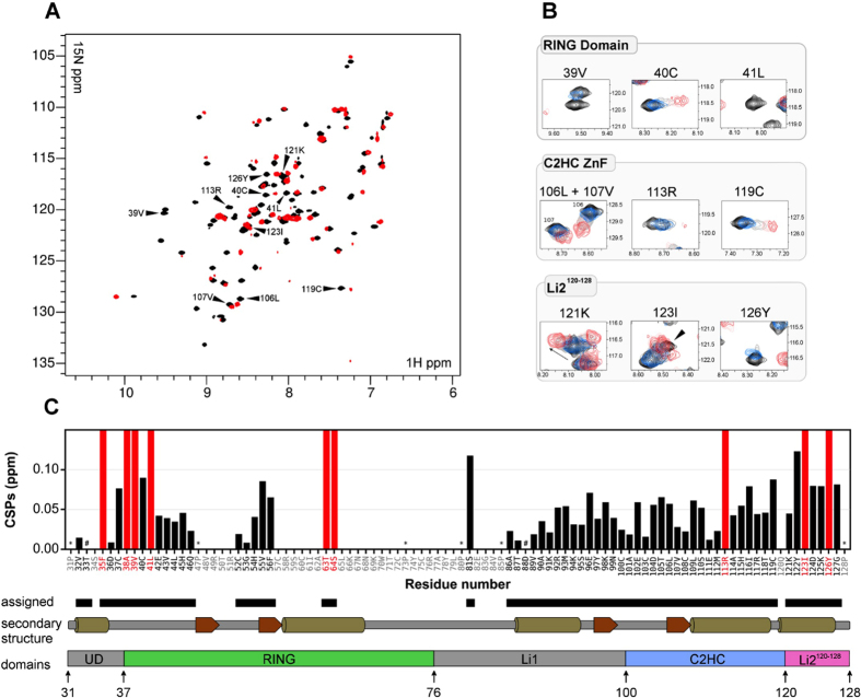
Figure 5. CSPs caused by UbcH5a in 15N-RNF125start31/stop129.
(A) Overlay of 1H-15N HSQC spectra of 0.2 mM 15N-RNF125start31/stop129 collected at 600 MHz before (black), and after addition of 2 molar equivalents of UbcH5a (red). Specific peaks shown in panel B are indicated by arrows. (B) Expanded regions of 1H-15N HSQC spectra showing specific residues in the RING domain, C2HC ZnF and Li2120-128, respectively. Overlays are shown of 0.2 mM 15N-RNF125start31/stop129 before (black), or after UbcH5a was added at a molar ratio of 0.25 (blue), 0.5 (grey), 1 (pink) or 2 (red). Some peaks (V39, L41, R113) broadened beyond detection at the higher molar ratios of UbcH5a. (C) A plot of the combined 1H and 15N chemical shift perturbations for 15N-RNF125start31/stop129 residues obtained at 2 molar equivalents of UbcH5a (see A). Assigned residues are in black in the sequence underneath the graph, Pro residues are indicated by (*). #indicate T33 and D88 that were too broad in our spectra to be included in the CSP calculation. Red bars represent residues that broadened beyond detection in the presence of UbcH5a. Diagrams of secondary structure elements (α-helices as cylinders, and β-sheets as arrows) and the RNF125stop129 domain organization are also shown.

