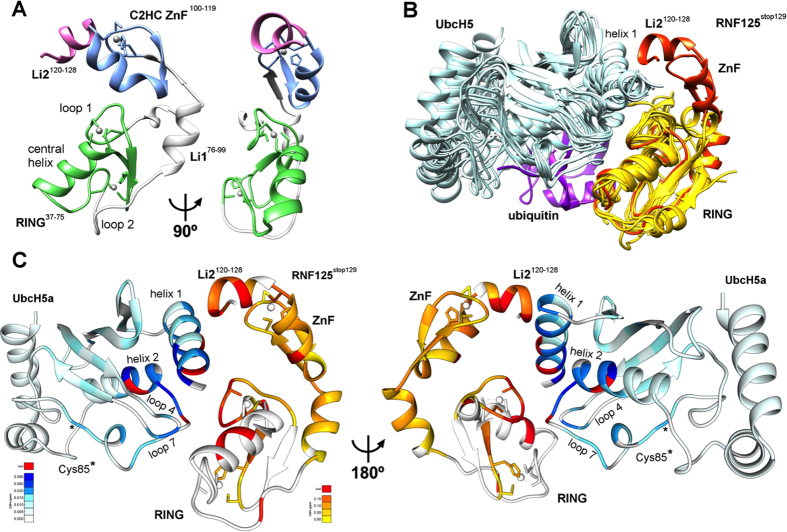
Figure 6. Crystal structure of RNF125stop129; models of UbcH5a interactions.
(A) Ribbon diagram of the RNF125stop129 structure with the RING domain in green, the C2HC ZnF in blue, the linker region L1 in grey and Li2120-128 in pink. A 90° rotated view is also shown. (B) Overlays of the RNF125stop129 structure (orange) with eight X-ray structures of RING domains (yellow) co-crystallized with UbcH5 E2 proteins (blue) or with a UbcH5a-Ub conjugate (4AP4). Ubiquitin is in purple. Structures from PDB were: 4QPL (RNF146/UbchH5a); 2YHO (Mylip/UbcH5a); 4V3K (RNF38/UbcH5b); 4AUQ (Birc7/UbcH5b); 4A4C (Cbl/UbcH5b); 3EB6 (IAP/ UbcH5b); 3RPG (Bmi/UbcH5c). Some PDB files were altered to depict the RING domain and E2 only. (C) Model of RNF125stop129 in association with UbcH5a, generated by overlaying RNF125stop129 with RNF146 in PDB 4QPL. The CSPs observed in the NMR experiments of Fig. 4 (15N-UbcH5a/RNF125start31/stop129 at 1:0.9) and Fig. 5 (15N-RNF125stop129/Ubch5a at 1:2) are highlighted on RNF125stop129 (yellow-orange) and UbcH5a (cyan-blue). Residues that broadened beyond detection are in red. A 180° rotated view is also shown.

