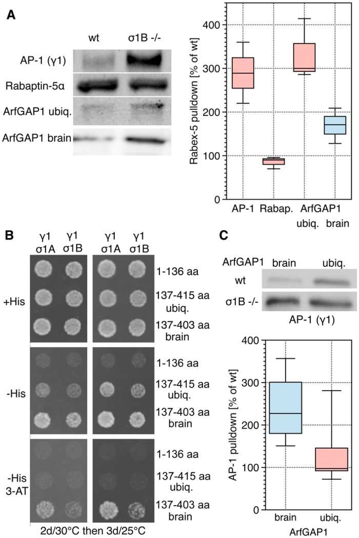
Figure 3. Specificity of AP-1 - ArfGAP1 complex formation.

Experiments were performed with extracts from individual animals. (A) Isolation of AP-1, Rabaptin-5α and ArfGAP1 isoforms from brain extracts by Rabex-5 pulldowns. Representative Western blots and the quantification (n = 3 each). (B) Y3H assay for ArfGAP1-σ1 binding specificities. The GAP-domain (aa 1–136) and the two C-terminal domains of ubiquitous (aa 137–415) and brain-specific ArfGAP1 (137–403) were tested. (C) ArfGAP1 isoforms pulldown AP-1 from wt and σ1B −/− brains. Representative Western blots and the quantification are shown (WB, br. n = 4 , ubiq. n = 5). Comparing the differences between ,ko‘ and wt (set to 100%) data sets gave in all χ2 test distributions ≤ 1 × 10−72 (>99% probability).
