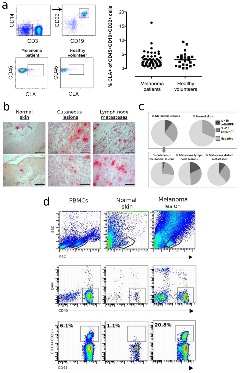
Figure 1. B cells may be recruited to skin and are present in melanoma lesions and normal skins.
(a) Proportion (%) of circulating B cells positive for the skin homing marker CLA from patients with melanoma (n = 49) and healthy volunteers (n = 24), quantified by multi-color flow cytometric evaluations. Peripheral blood B cells were identified as CD3-CD14-CD19+CCD22+ cells (top panel). Representative dot plots of CLA+CD45+B cells from a melanoma patient and a healthy volunteer are shown (bottom panel). Quantification was based on % CLA+B cells from CD45+CD3-CD14-CD19+CCD22+PBMCs (right panel). (b) CD22+ cells in normal skin (left), cutaneous melanoma lesions (middle) and lymph node metastases (right) were detected by immunohistochemistry (tissue microarrays, top/bottom: example images per cohort, acquired on a Leica AxioScan, 40x objective; Scale bars: 66 μm). (c) CD22+ cell infiltrates per high-powered field (HPF) were quantified; Top panel: n = 189 melanomas, n = 16 normal skin samples; Bottom panel: CD22+ infiltrates stratified into cutaneous, lymph node and distant metastases. (d) B cells were detected from peripheral blood, melanoma lesion and normal skin samples using antibodies against CD19+, CD22+ (both FITC) and CD45+ (PE) and analysed by flow cytometry (example from matched blood, normal skin and cutaneous melanoma samples derived from a patient with melanoma).

