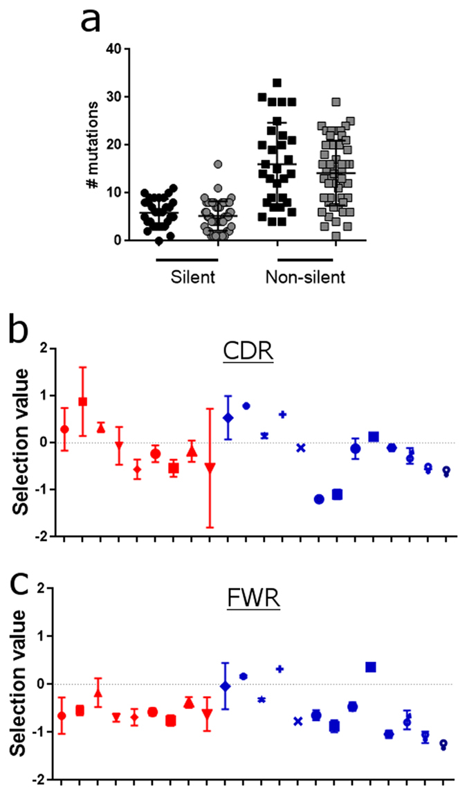
Figure 4. Similar mutational rates and clonal selection in melanoma and normal skin IgG antibodies.

(a) Silent and non-silent mutations in the IgG VH regions determined using IMGT V-quest. Healthy volunteer skin (grey) and melanoma (black) samples show no significant differences in the antibody mutational rates. (b&c) Quantified clonal selection in the CDR (b) and FWR (c) regions from IgG VH sequences for each melanoma (red symbols ± SEM) and normal skin (blue symbols ± SEM) samples. NS: not significant. Please see Supplementary Materials and Methods (each symbol represents the mean value of selection from the sequences from each individual donor).
