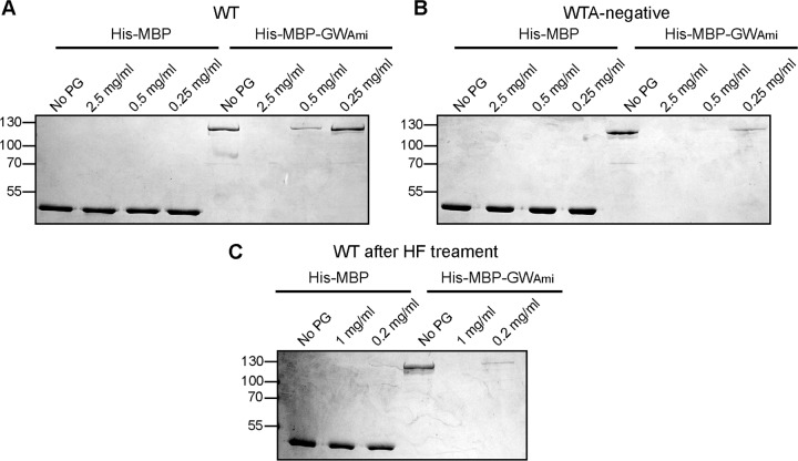
FIG 9.
Binding of His-MBP-GWAmi to purified peptidoglycan isolated from wild-type or WTA-deficient L. monocytogenes cells. (A and B) Peptidoglycan isolated from strain 10403S (WT) or the WTA-deficient strain 10403S ΔtagO1-2sup (WTA negative) was suspended at the indicated concentration prior to HF treatment and incubated with purified His-MBP or His-MBP-GWAmi protein. “No PG” indicates control samples where the proteins were incubated without peptidoglycan. Peptidoglycan and bound protein were removed by centrifugation, an aliquot of the supernatant fraction was analyzed by SDS-PAGE, and the proteins were visualized by Coomassie staining. (C) Peptidoglycan isolated from strain 10403S (WT) was suspended at the indicated concentration after HF treatment to chemically remove WTA and incubated with purified His-MBP or His-MBP-GWAmi protein and processed as described above. The His-MBP or His-MBP-GWAmi proteins have predicted sizes of 45 kDa and 118 kDa, respectively. The positions of protein molecular mass markers are indicated on the left in kilodaltons. Representative gels from three independent experiments are shown.

