FIG 6.
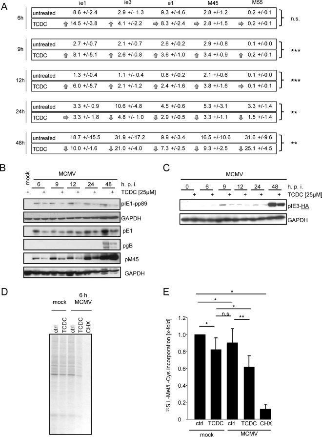
TCDC inhibits MCMV replication at the level of protein translation and modifies viral mRNA expression. (A) Primary mouse hepatocytes were incubated with 25 μM TCDC for 3 h and infected with Δm157-MCMV-luciferase afterwards. Cells were lysed after 6 h, 9 h, 12 h, 24 h, or 48 h, and RNA was isolated. cDNA was synthesized, and quantitative PCR was performed with specific primers for ie1, ie3, early1, M45, and M55. Depicted are the relative RNA expression levels in comparison to that of the cellular transcript sdha. To calculate statistical significance, the results of all transcription values at one time point were considered (except in case of M55, where no gene expression was detectable in the 6- to 12-h period and only later time points were included in the analysis). (B) Primary hepatocytes were treated for 3 h with 25 μM TCDC and infected with MCMV-IE3-HA or Δm157-MCMV-luciferase (multiplicity of infection of 0.1). Hepatocytes were lysed at the indicated time points and pIE1, pIE3-HA, pE1, pM45, gB, and housekeeping GAPDH were detected by SDS-PAGE and immunoblotting with specific antibodies. (C) Primary mouse hepatocytes were preincubated for 3 h with 25 μM TCDC and infected with MCMV (1 PFU/cell). 35S-labeled l-Met/l-Cys (27 μCi) was added to the cells directly after centrifugally enhanced infection. After 6 h, cells were lysed with lysis buffer, proteins were separated by SDS-PAGE, the gel was fixed and dried, and the intensity of radioactively labeled proteins was visualized by autoradiography. The inhibitor of translation CHX was used as a positive control. (D) Intensities of 35S incorporation were quantified (for details, see Materials and Methods). The intensities were calculated in relation to untreated control samples.

