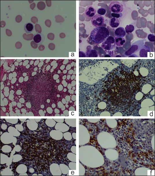
Figure 1.

Peripheral blood smear shows large granular lymphocyte (a) [100x, May-Grünwald-Giemsa (MGG) stain]; Bone marrow aspirate shows erythroblastopenia and large granular lymphocytes (b) (100x, MGG stain); trephine biopsy shows reactive lymphoid nodules (c) (20x, H&E stain) composed of CD20+ cells (d) (20x) and CD3+ cells (e) (20x); CD8+ cells in the interstitium (f) (20x)
