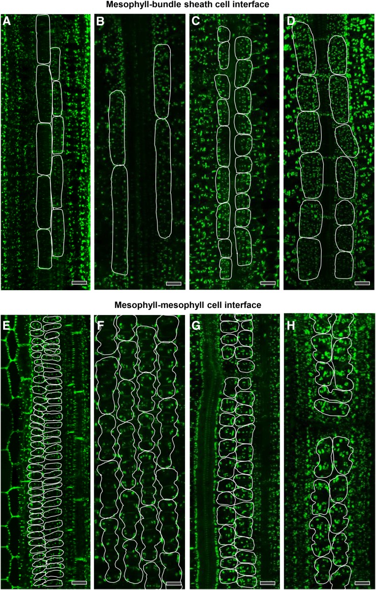
Figure 8.
Pitfield Distribution at Cell Interfaces in Leaves of C3 and C4 Species after Immunofluorescence Detection of β-1,3-Glucan.
(A) and (E) Rice, C3
(B) and (F) Wheat, C3.
(C) and (G) S. viridis, C4.
(D) and (H) Maize, C4.
Pitfields are in green (Alexa Fluor 488 fluorescence). In (A) to (D), bundle sheath cell surface areas in direct contact with mesophyll cells are outlined in white. In (E) to (H), mesophyll cell surface areas in direct contact with other mesophyll cells are outlined in white. Bars = 20 µm.

