Fig. 1.

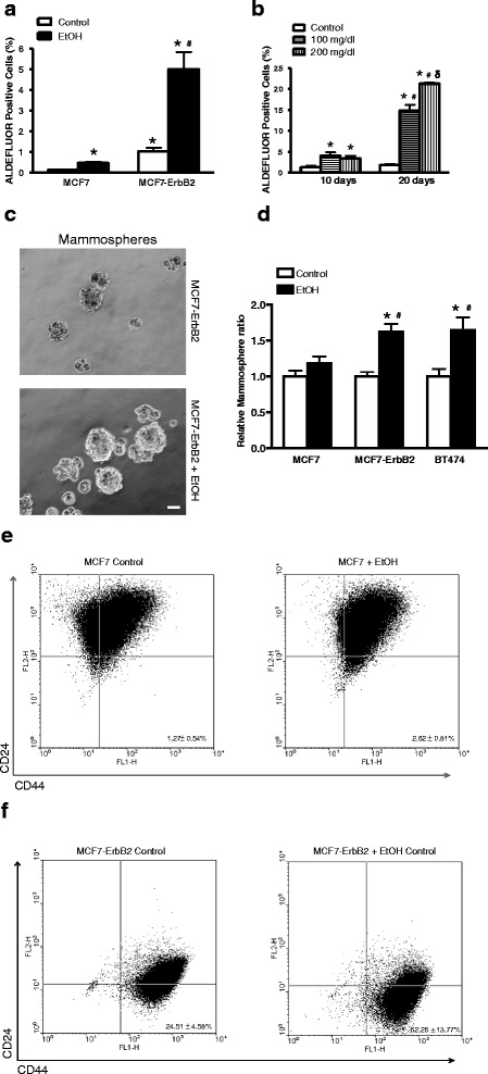
Effect of alcohol on cancer stem-like cell (CSC) population. a MCF7 or MCF7-ErbB2 cells were exposed to alcohol (0 or 100 mg/dl) for 10 days, and then were processed for ALDEFLUOR assay, followed by flow cytometry for the detection of CSCs as described in the Materials and Methods. CSC population was calculated as percentage of total cells population. Each data point was mean ± SEM of three independent experiments. *denotes significant difference from respective control groups. #denotes significant difference from alcohol-treated MCF7 cells. b MCF-ErbB2 cells were exposed to alcohol (0, 100 or 200 mg/dl) for 10 or 20 days and then CSC population was determined as described above. *denotes significant difference from respective control groups. #denotes significant difference from respective 10 day-alcohol-exposed groups. δ denotes significant difference from 100 mg/dl alcohol-exposed groups during the 20 day exposure period. c and d MCF7, MCF7-ErbB2 or BT474 cells were exposed to alcohol (0 or 100 mg/dl) for 10 days, then 1000 cells were cultured on ultra-low attachment plates for assaying mammosphere formation as described in the Materials and Methods. The cell morphology was captured by a Zeiss Axiovert 40C photomicroscope. The number of mammospheres was determined. Each data point was the mean ± SEM of three independent experiments. *denotes significant difference from respective control groups. #denotes significant difference from alcohol-treated MCF7 cells. Bar = 50 μm. e and f Expression of breast cancer stem cell markers CD44+/CD24− in MCF7 (e) or MCF7-ErbB2 (f) cells treated with or without Ethanol (100 mg/dl) was determined by flow cytometry. All experiments were repeated at least three times and there were triplicates for each replication. Each data point was the mean ± SD of three independent experiments
