
Fig. 1.
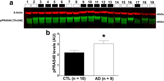
Phosphorylated PRAS40 levels are increased in AD patients. a Western blots of proteins extracted from the inferior temporal gyrus of human control (CTL; n = 10) and AD brains (n = 9). Blots were probed with the indicated antibodies. The descriptive information for each patient (numbered above the blots) are reported in Table 1. b Quantitative analysis of the blot shows that phosphorylated PRAS40 levels were significantly higher in AD compared to CTL cases (p = 0.0325). Quantitative analyses of the blots were obtained by normalizing PRAS40 levels to β-Actin, used as a loading control. Error bars represent mean ± SEM