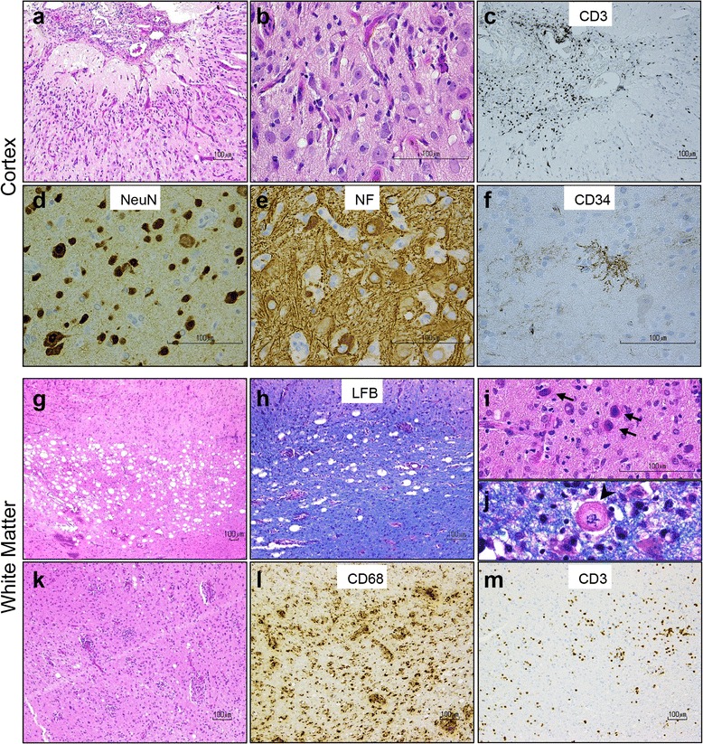
Fig. 2.

Histological analysis of right occipital brain biopsy reveals unusual form of inflammatory encephalopathy. a-b Hematoxylin and eosin (H&E) stains show chronic meningitis and neocortex with architectural neuronal disorganization, associated with reactive vasculature, gliosis, microglial activation, and mild chronic inflammation. c CD3+ T-lymphocytes predominate in the meninges. d Many cortical neurons display dysmorphic features, such as increased size, displaced Nissl substance, clustering, and e abnormal phosphorylated neurofilament accumulation in their cell body. f Tuft-like ramified CD34-positive processes are also seen in the neocortex. g-j The subcortical white matter contains multifocal vacuolization associated with minimal myelin loss on luxol fast blue (LFB) stain, and scattered bizarre oligodendroglial-like cells with enlarged nuclei containing glassy, homogeneous nuclear inclusions (i, arrows) and Creutzfeldt-like astrocytes (j, arrowhead). k-m Prominent chronic perivascular and intraparenchymal inflammatory infiltrate in the white matter is composed of CD68+ macrophage/microglia and CD3+ T-lymphocytes
