Fig. 5.

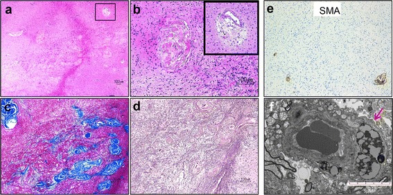
Unusual form of sclerosing vasculopathy in HED-ID. a-b Hematoxylin and eosin stain shows vessels in the white matter with variably obliterated lumina, hyalinization, and perivascular hemosiderin deposits (inset in b). c-d Concentric lamellated pattern of collagen accumulation is seen on trichrome (c), extending into the adjacent brain parenchyma and forming a reticulin-rich scar (d). e Sclerotic vessels lack smooth muscle fibers in their walls. f EM shows capillary vessel with aberrant accumulation of lysosomes (arrow); scale bar = 5 μm
