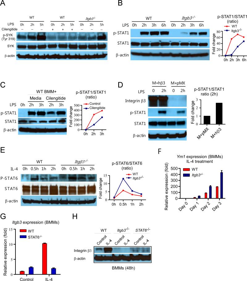
Figure 5. Integrin beta3 signaling favors STAT1 activation and suppression of STAT6 signaling in macrophages.
(A) Western blots of p-SYK and SYK in LPS-stimulated WT and Itgb3−/− BMMs. (B) Western blots of p-STAT1 and STAT1 in LPS-stimulated WT and Itgb3−/− BMMs. (C) Western blots of p-STAT1 and STAT1 in LPS-stimulated WT BMMs with or without cilengitide pretreatment. (D) Western blots of p-STAT1 and total STAT1 in WT BMMs overexpressing integrin beta3. M+pMX: macrophage treated with empty pMX vector. M+hβ3: macrophage treated with human integrin beta3 constructed pMX vector. (E) Western blots of p-STAT6 and total STAT6 in IL-4 stimulated WT and Itgb3−/− BMMs. (F) Ym1 mRNA expression after IL-4 treatment of WT and Itgb3−/− BMMs. (G) Itgb3 mRNA expression level after IL-4 treatment in Stat6−/− macrophages. (H) Western blot analysis of integrin beta3 expression in WT, Itgb3−/−, and Stat6−/− BMMs after 5ng/ml IL-4 treatment for 48 hours.

