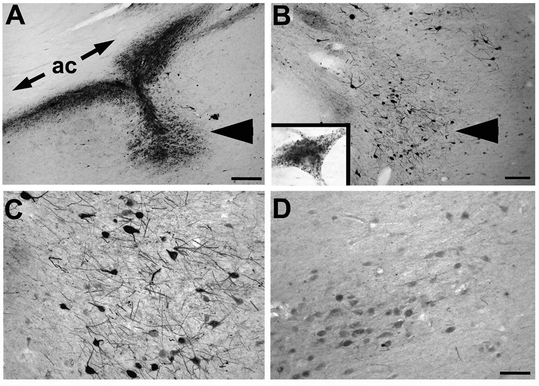
Figure 2. AAV2-NGF gene expression.
(A) NGF labeling shows site of NGF gene delivery in nucleus basalis of Meynert (arrowhead), under anterior commissure (ac). Patient injected three years previously. (B) Another site from same brain showing NGF uptake in neurons in region of nucleus basalis (arrowhead). Inset shows single neuron with granular intraneuronal labeling. (C) Higher magnification of NGF-expressing neurons, compared to (D) less intense labeling in nucleus basalis neurons located 3mm from injection site. Scale bar A 325µm; B 250µm; C–D, 100µm.

