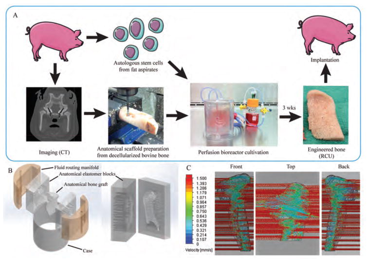
Figure 1. Fabrication of engineered bone grafts.
(A) Personalized bone tissue engineering process. Autologous mesenchymal stem cells (from fat aspirates) and CT images were obtained for each animal subject. The anatomical scaffold was fabricated from the bovine stifle bone that had been processed to remove any cellular material while preserving the tissue matrix. The cells were seeded into the scaffold and cultured in the specially designed perfusion bioreactor. After 3 weeks, the engineered RCU was implanted back into the pig for 6 months. (B) The bioreactor culture chamber consisted of five components designed to provide tightly controlled perfusion through the anatomical scaffold: anatomical inner chamber with bone scaffold; two halves of the polydimethylsiloxane (PDMS) (elastomer) block with incorporated channels; two manifolds; and an outer casing. The PDMS block was designed as an impression of the anatomical RCU structure, and contained flow channels at both sides for the flow of culture medium in and out of the scaffold. The flow rate necessary for providing nutrient supply and hydrodynamic shear was defined by the design of parallel channels in the elastomer block (with the channel diameters and distribution dictated by the geometry of the graft) and the fluid routing manifold. The channel diameters and spacing were specifically designed by flow simulation software for each pig, to provide a desired interstitial flow velocity for a given shape and size of the anatomical RCU. (C) Flow simulation of the medium flow through the anatomically shaped scaffold reveals uniform flow velocity throughout the volume of the graft.

