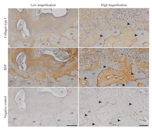
Figure 6. Deposition of type I collagen and bone sialoprotein (BSP) in the newly formed bone.
The regenerating new bone (nb) within the tissue-engineered bone graft (g) is shown at low and high magnification. Localized areas of type I collagen and BSP at the graft-host bone interface were precursors for bone formation. New bone contained osteocytes (black arrowheads) within their lacunae. Negative immunohistochemistry confirmed the specificity of the staining. Images are representative of n=4 animals. Scale bars: 400 μm (low) and 100 μm (high).

