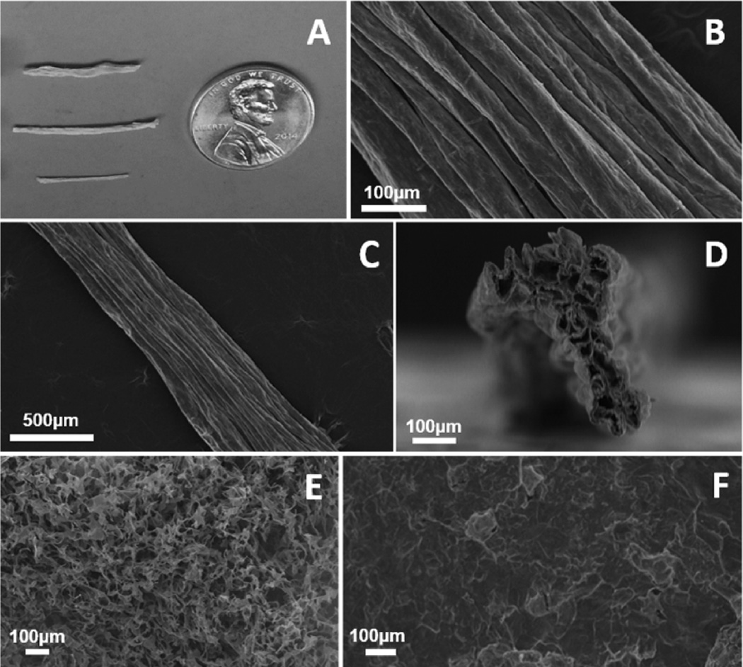
Figure 1.
Example images of collagen scaffold morphology. (A) Optical image of scaffolds fabricated of various diameters (top to bottom 6350 µm, 4762 µm, and 3175 µm). (B) SEM revealed highly aligned topographical features along the surface of collagen scaffolds. (C) Aligned features were maintained uniformly along the length of collagen scaffolds. (D) Cross sectional SEM images revealed a highly porous interior within the scaffolds. (E) Scaffolds fabricated from unassembled collagen suspensions and (F) scaffolds fabricated from fibrillar collagen in low aspect ratio vessels showed no preferential orientation of topographical features. All samples featured here were fabricated at a collagen concentration of 2.0 mg/mL and a freezing temperature of −80 °C and were imaged in the dry, as-fabricated state.

