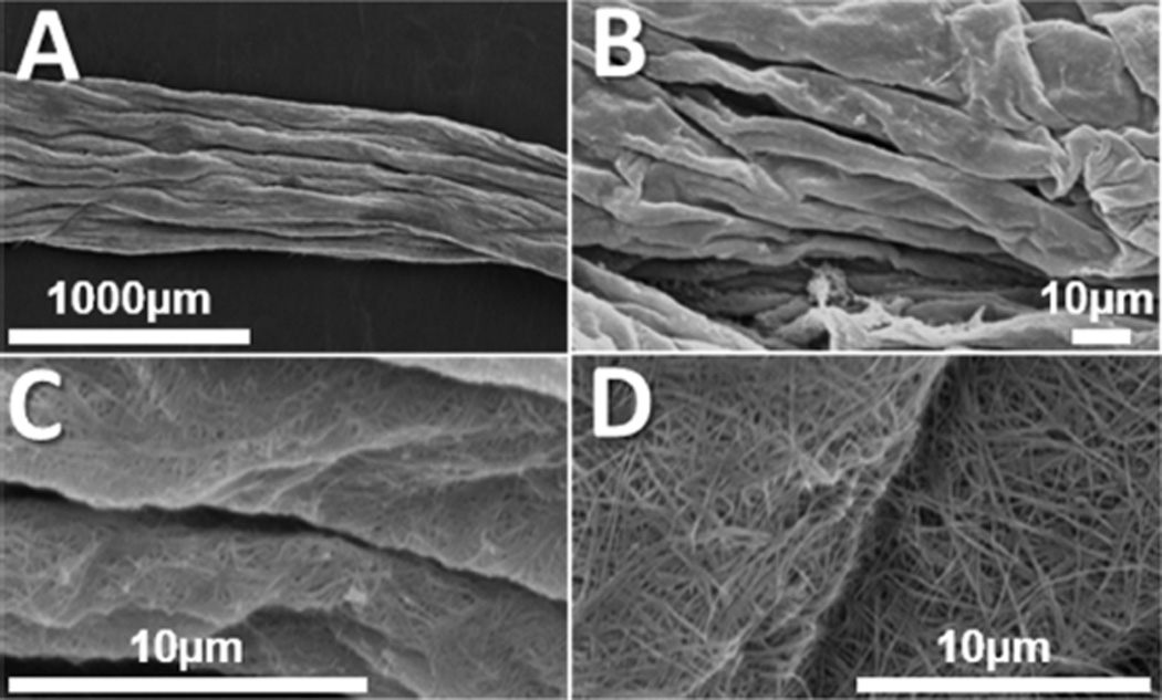
Figure 4.
Representative SEM images of aligned collagen scaffolds critical point dried in the hydrated state. (A) Aligned topographical features are preserved in the hydrated state observed at 50×. (B) Folds and ridges of individual features are aligned in the same direction as other scaffold features observed at 1300×. (C) The surface of collagen scaffolds shows evidence of a randomly oriented, fibrillar topography at high magnification. Panels B and C are high magnification frames of panel A. (D) Representative image showing randomly oriented, fibrillar topography on the surface of a separate collagen scaffold.

