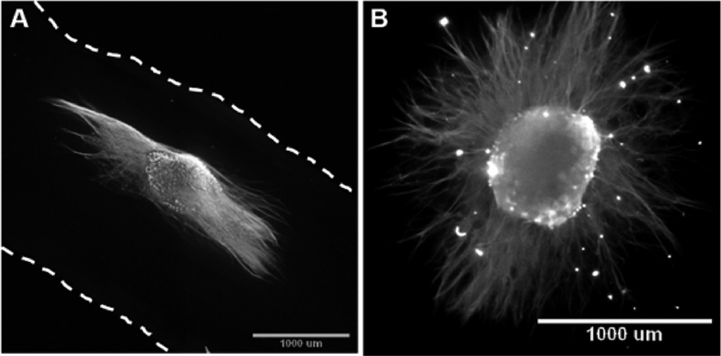
Figure 7.
Chick dorsal root ganglia explants cultured on both aligned and randomly oriented collagen scaffolds and immunolabeled with antineurofilament M. (A) Epifluorescence image of DRG extending axons along the length of aligned collagen scaffold (dashed lines indicate the edge of scaffold). (B) Epifluoresence image of DRG explant cultured on a randomly oriented collagen scaffold fabricated in a low aspect ratio vessel, which demonstrated no preferentially oriented outgrowth.

