
Figure 5.
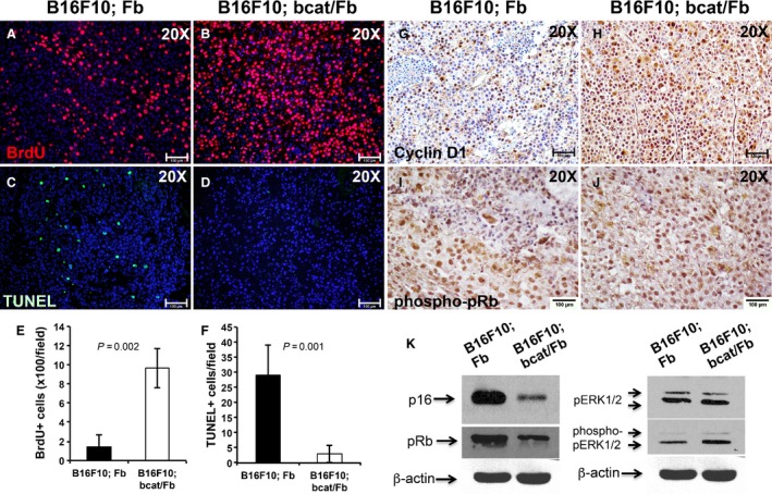
Dermal fibroblasts suppress early stage melanoma tumor growth by inducing a cell cycle arrest in tumor cells. Cell proliferation in control (A) and mutant tumors (B) was examined by BrdU labeling and immunostaining. To detect cell apoptosis in control (C) and mutant (D) tumors, TUNEL staining was performed. (E) Number of BrdU positive cells per field (20×) is showed as the mean ± SEM, n = 3. (F) Number of TUNEL positive cells per field (20×) is showed as the mean ± SEM, n = 3. Reduced immunostaining of Cyclin D1 was detected in B16F10; Fb tumor (G) as compared with B16F10; bcat/Fb tumor sample (H). phospho‐pRb expression was evaluated on paraffin sections of B16F10; Fb tumor (I) and B16; bcat/Fb tumor (J) by immunohistochemistry. Images shown are representative of 5 pairs of melanoma samples. (K) Expression of p16, pRb, pERK1/2, and phospho‐pERK1/2 protein was analyzed and compared between B16F10; Fb and B16; bcat/Fb tumors by western blotting. β‐actin was used as internal control. Data are representative of three independent experiments. Scale bar: A–D and G–J,100 μm.