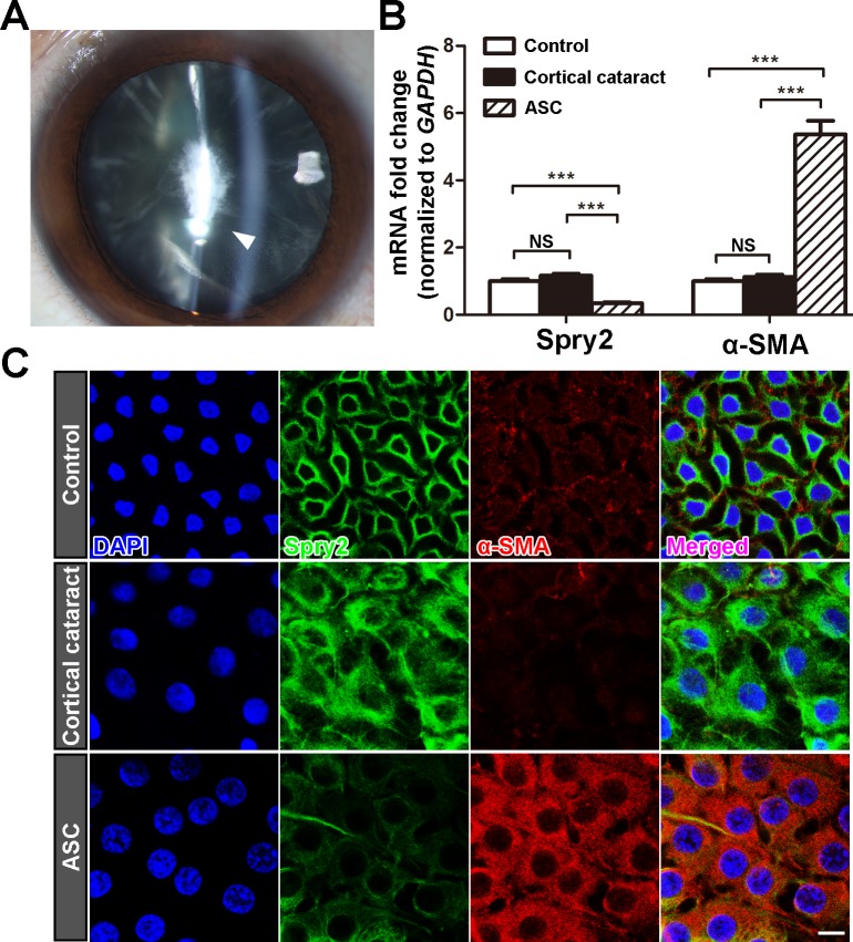
Fig 1. Spry2 expression level is decreased in anterior capsule LECs of ASC patients.
(A) Representative slit-lamp microscope photo of an ASC patient (60 years old, Female). The white arrow indicates the irregular fibrotic opacity beneath the anterior capsule. (B) Total RNA was extracted from the anterior capsules of ASC patients, age-matched cortical cataract patients and postmortem human lens (control). The mRNA level of Spry2 was determined using real-time PCR and normalized to GAPDH. ***P<0.001, NS: not significant, n = 6. Fold change relative to the level of the control groups is displayed. (C) Lens anterior capsule whole-mounts from ASC patients, age-matched cortical cataract patients, and postmortem human lens (control) were probed for Spry2 (green), α-SMA (red) and DAPI (blue). Images were acquired from the central area of each sample. Scale bar: 10μm.

