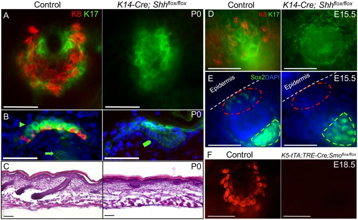
Fig 4. Intraepithelial Shh signaling is required for TD MC development.
(A) K8 and K17 whole mount staining in control and K14-Cre; Shhflox/flox skin at P0. (B) K8 and K17 section staining in control and K14-Cre; Shhflox/flox skin at P0. Arrowhead, K17+ TD keratinocytes. Arrow, K17+ hair follicle. Pentagon, abortive K17+ hair germ. (C) H&E staining in sections of control and K14-Cre; Shhflox/flox skin at P0. (D) K8 and K17 whole mount staining in control and K14-Cre; Shhflox/flox skin at E15.5. (E) Confocal maximum projection oblique view of Sox2 whole mount staining in control and K14-Cre; Shhflox/flox skin at E15.5. Red circle, epidermal surface over primary hair germ. Green outline, dermal papilla. (F) K8 whole mount staining in control and K5-tTA; TRE-Cre; Smoflox/flox skin at E18.5. Scale bars, 50 μm.

