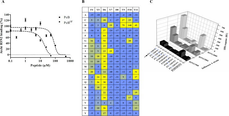
Fig 6. Enhancement of peptide binding affinity to HA by site-directed substitution.
A) Inhibition of Aichi H3N2 binding to immobilized fetuin by PeB-Lys and PeBGF-Lys as determined via SPR (n = 2). Error bars indicate the SEM. Dashed lines represent a four parameter logistic fit. B) Microarray based substitutional analysis of PeB using fluorescently labeled Aichi H3N2 viruses as analytes (for further virus strains see S7 Fig). Amino acids in the first row represent that of PeB, while amino acids in the first column show the substitution. The values represent the contrast relative to fetuin as positive control (see Material and Methods). C) Binding of different viruses to selected immobilized single and double amino acid substituted variants of peptide PeB-Lys. The binding response from SPR measurements is shown (as in Fig 3).

