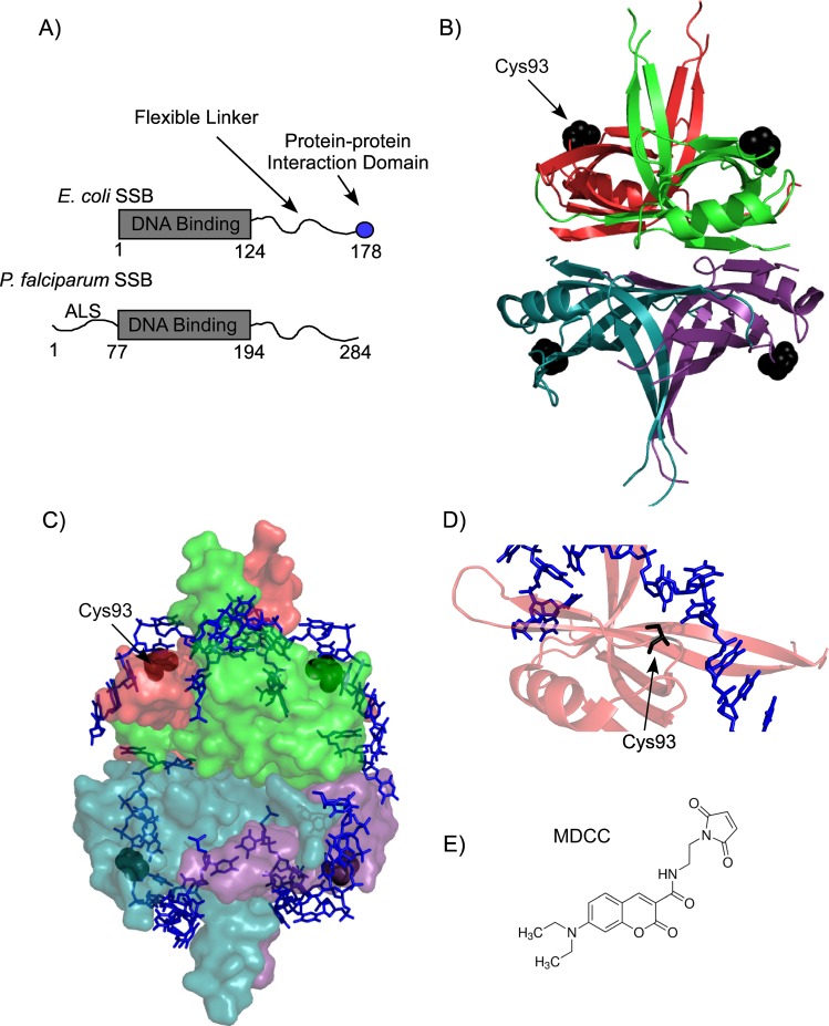
Fig 1. Domain organization of Pf-SSB.
(A) Schematic representation of the DNA binding, protein-protein interaction and linker regions of E. coli and P. falciparum SSB. Pf-SSB also has an apicoplast localization signal (ALS), which is not required for its DNA binding function. The numbers denote positions of the amino acids at the beginning and end of each domain. (B) Individual subunits of the homotetrameric DNA binding domain are depicted as cartoon representation in the crystal structure of Pf-SSB. The Cys93 residues used for attachment of the fluorophore are shown as black spheres. (C) Pf-SSB is shown as surface representation with two (dT)35 DNA molecules (blue stick representation) wrapped around the homotetramer. (D) The proximity of Cys93 (black stick) to the bound DNA in Pf-SSB is highlighted. (E) Structure of the MDCC (7-diethylamino-3-((((2-maleimidyl)ethyl)amino)-carbonyl)coumarin) fluorophore used to label Pf-SSB. Images of the Pf-SSB structure were generated using PDB ID: 3ULP.

