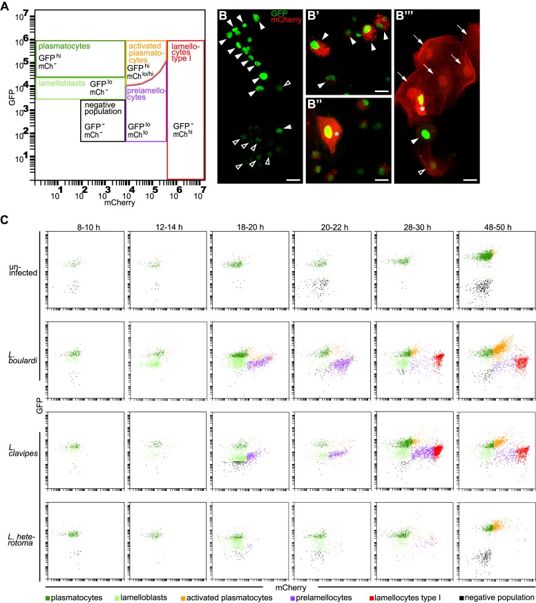
Fig 1. Six different hemocyte classes.
(A) Hemocyte gates. Plasmatocytes (GFPhimCh-) and lamelloblasts (GFPlomCh-) expressed only GFP. Activated plasmatocytes (GFPhimChlo) were equal in size to plasmatocytes and had varying amounts of mCherry-positive punctae in their cytoplasm. Similar to activated plasmatocytes, type II lamellocytes expressed both markers (GFPhi mChhi). Type II lamellocytes were larger than plasmatocytes, similar in shape to lamellocytes, and with strong expression of mCherry in their cytoplasm. Prelamellocytes (GFPlomChlo) had low GFP expression while increasing their mCherry expression. Type I lamellocytes (GFP-mChhi) expressed only mCherry. The negative population expressed neither GFP nor mCherry. (B-B”‘) Representative images of hemocyte types. (B) plasmatocytes (filled arrowheads) and lamelloblasts (open arrowheads). (B’) Activated plasmatocytes (filled arrowheads). (B”) Type II lamellocyte (star). (B”‘) Lamellocytes type I (arrows), prelamellocyte (open arrowhead), activated plasmatocyte (filled arrowhead), and lamellocyte type II (star). Scale bars 10 μm. (C) Flow cytometry plots at representative time points after a wasp infection. Me/w larvae were uninfected or infected by L. boulardi, L. clavipes, and L. heterotoma. The time points were chosen to cover major changes in the composition of the hemocyte population in the course of the time line experiment. In uninfected animals mainly plasmatocytes were present in the circulation. The cellular immune response after infection by L. boulardi and L. clavipes proceeded in a stereotypical way. At 10–12 h after infection, two plasmatocyte-like populations, plasmatocytes and lamelloblasts, were present in the circulation. At 18–20 h after infection, lamelloblasts developed into prelamellocytes. Plasmatocytes started to appear already 8–10 h after infection, and were the dominant cell type at 48–50 h. The first type I lamellocytes were seen in the circulation 20–22 h after infection. Large numbers of type I lamellocytes were in the circulation 28–30 h and 48–50 h after infection. A L. heterotoma infection induced a similar immune response until 18–20 h after infection. Then the numbers of prelamellocytes and lamellocytes type I were reduced in comparison to L. boulardi and L. clavipes-infected larvae. 48–50 h after a L. heterotoma infection, plasmatocytes and activated plasmatocytes were the dominant hemocyte types present. They were accompanied by only very few type I lamellocytes.

